Every living cell exists in a state of perpetual negotiation with its environment. From fluctuating temperatures and oxygen deprivation to oxidative damage and inflammation, the biological landscape is unrelenting in its demands. Yet remarkably, cells have evolved a sophisticated molecular defence system capable of responding to these threats within minutes – not days. At the centre of this system are heat shock proteins (HSPs): ancient, conserved, and extraordinarily precise molecular guardians that stand between cellular integrity and catastrophic protein collapse. Understanding stress and heat shock proteins, and the cellular protection they confer, is not merely an academic exercise – it is foundational to understanding how life persists under pressure.
What Are Heat Shock Proteins, and How Were They First Discovered?
The story of heat shock proteins begins in 1962, when Italian geneticist Ferruccio Ritossa observed an unusual chromosomal “puffing” pattern in fruit fly (Drosophila) chromosomes that had been exposed to elevated temperatures. What he had inadvertently witnessed was one of biology’s most fundamental survival responses – a cellular alarm system triggered by stress.
Heat shock proteins are a highly conserved family of proteins produced across virtually all living organisms, from bacteria to human beings. Their evolutionary persistence across billions of years underscores their critical importance: these molecules are not optional cellular accessories, but essential survival infrastructure.
Under baseline physiological conditions, HSPs constitute approximately 5–10% of total cellular protein content. However, when a cell encounters stress – whether thermal, chemical, hypoxic, or inflammatory – the heat shock response (HSR) is rapidly activated, potentially elevating HSP concentration to 15–25% of total intracellular protein within minutes.
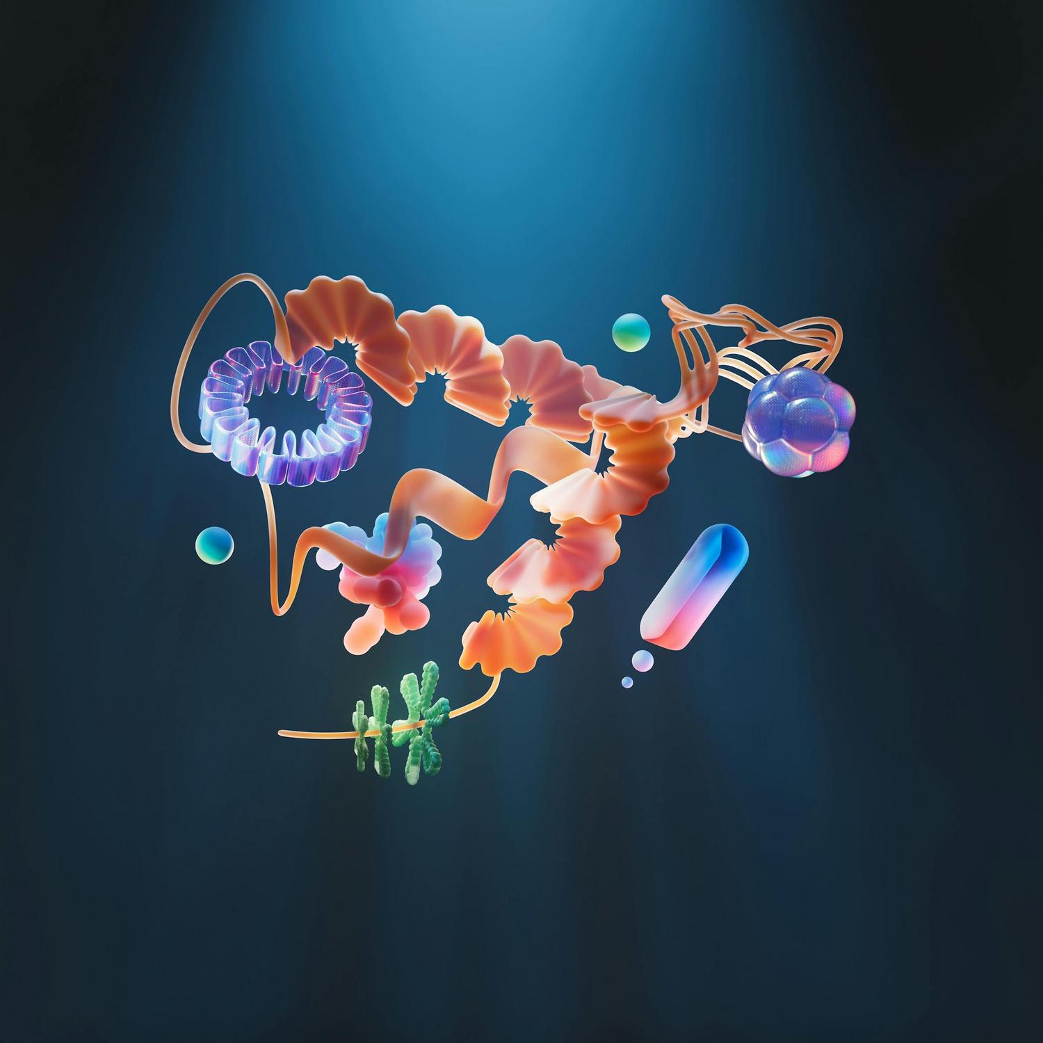
HSPs are classified primarily according to their molecular weight (measured in kilodaltons, kDa), with each family exhibiting distinct structural characteristics and protective roles. The table below summarises the principal HSP families and their key functions.
HSP Family Classifications and Cellular Roles
| HSP Family | Molecular Weight | Primary Location | Key Function |
|---|---|---|---|
| HSP100 | 104–110 kDa | Cytosol, Nucleus | Protein disaggregation; degradation under severe stress |
| HSP90 | ~90 kDa | Cytosol | Stabilises substrate proteins; steroid receptor complex; ATP-dependent |
| HSP70 | ~72 kDa | Cytosol, Nucleus, ER, Mitochondria | Highly stress-inducible; protein folding/unfolding; thermotolerance |
| HSC70 | ~73 kDa | Cytosol | Constitutively expressed; protein transport to organelles |
| HSP60 | ~60 kDa | Mitochondria | Chaperonin function; maintains respiratory-chain integrity |
| HSP40 | ~40 kDa | Multiple | Co-chaperone; works with HSP70; stress-responsive |
| Small HSPs (sHSPs) | 12–43 kDa | Cytosol, Nucleus | ATP-independent; holdases and sequesterases; antioxidant properties |
How Do Heat Shock Proteins Provide Cellular Protection During Stress?
The protective capacity of heat shock proteins rests upon their function as molecular chaperones – proteins that supervise the structural integrity of other proteins throughout their lifecycle. This chaperone activity is multidimensional and operates through several coordinated mechanisms.
Protein Folding and Refolding
Newly synthesised polypeptides are vulnerable to misfolding, particularly under stressful conditions. HSPs bind to exposed hydrophobic regions of these nascent proteins, stabilising them and guiding them towards their correct three-dimensional configurations. When proteins have already been damaged or partially denatured by stress, HSPs facilitate their refolding back to functional states – a recovery process that would otherwise be impossible.
Prevention of Protein Aggregation
Misfolded proteins have a strong tendency to clump together, forming aggregates that are not only non-functional but also cytotoxic. This aggregation is a hallmark of numerous degenerative diseases. HSPs – particularly small HSPs such as HSP27 – act as holdases and sequesterases, physically preventing irreversible aggregation and preserving the solubility of damaged proteins until conditions permit their proper refolding or degradation.
Protein Quality Control and Degradation
Not all damaged proteins can be salvaged. HSPs participate in directing irreversibly compromised proteins toward the ubiquitin-proteasome system (UPS) or autophagy-lysosome pathway for controlled degradation. This quality control function is essential for maintaining cellular cleanliness – a process collectively known as proteostasis (protein homeostasis).
Thermotolerance and Cross-Tolerance
A particularly elegant property of the heat shock response is its capacity to confer thermotolerance: prior exposure to a sublethal stressor transiently equips cells to withstand subsequent exposures that would otherwise be lethal. Moreover, this protection extends across different stress modalities – a phenomenon termed “cross-tolerance” – providing cellular resilience against simulated hypoxia and ischaemia, not merely heat.
How Is the Heat Shock Response Regulated at a Molecular Level?
The orchestration of the heat shock response is governed by a family of transcription factors known as heat shock factors (HSFs), of which HSF1 is the primary stress-activated regulator in human cells.
Under normal conditions, HSF1 exists in an inactive, monomeric form, held in complex with HSP90 and co-chaperones including FKBP52. When cellular stress causes an accumulation of misfolded proteins, these proteins compete for and sequester HSP90, liberating HSF1. Free HSF1 monomers then oligomerise into active trimers, translocate to the nucleus, and bind to specific DNA sequences – heat shock elements (HSEs) – within the promoter regions of HSP genes.
This binding triggers the rapid transcription and translation of HSPs, dramatically amplifying the cell’s protective capacity within minutes. Once cellular stress subsides and proteostasis is restored, HSP70 and HSP90 bind directly to HSF1 trimers, creating a negative feedback loop that attenuates the response. HSP mRNA is subsequently degraded at an accelerated rate, allowing the system to reset.
HSF2 provides a complementary regulatory layer, supporting HSF1 during acute stress whilst also playing roles in cellular development and differentiation under normal conditions.
How Do Stress and Heat Shock Proteins Defend Against Oxidative Damage and Programmed Cell Death?
Among the most clinically significant dimensions of heat shock protein biology is the relationship between stress, heat shock proteins, and cellular protection from oxidative injury and apoptosis (programmed cell death).
Oxidative Stress Defence
Reactive oxygen species (ROS) are generated as natural byproducts of cellular metabolism, but their accumulation under stress conditions causes profound molecular damage. HSP70 counteracts oxidative stress through multiple simultaneous mechanisms: inhibiting the aggregation of oxidatively damaged proteins, facilitating their clearance via the 20S proteasome, inducing expression of antioxidant enzymes including superoxide dismutase (SOD1) and catalase, and mobilising the cellular glutathione system – a critical intracellular antioxidant network – by increasing both cytosolic and mitochondrial reduced glutathione pools.
HSP27 demonstrates particularly potent antioxidant properties. Large oligomers of this small HSP maintain glutathione in its reduced (active) form and have been shown to abolish lethal intracellular ROS bursts, providing especially strong cytoprotection in neuronal cells. Furthermore, oxidative stress activates the redox-sensitive transcription factor Nrf2 in concert with HSFs, inducing a coordinated cytoprotective gene expression programme.
Anti-Apoptotic Activity
Apoptosis, while a necessary process for development and tissue maintenance, becomes pathological when inappropriately activated under stress. HSPs interrupt apoptotic cascades at multiple intervention points.
HSP70 blocks the mitochondrial apoptotic pathway by preventing the release of cytochrome c, inhibiting the transition of procaspase-9 to its active form, and blocking the translocation of apoptosis-inducing factor (AIF) to the nucleus. It simultaneously inhibits receptor-mediated death pathways, including those triggered by TNFα and Fas ligand signalling.
HSP27, meanwhile, demonstrates broad anti-apoptotic activity by interacting directly with procaspase-9 and procaspase-3, preventing their upstream cleavage and thus interrupting apoptotic signalling before it reaches its most damaging stages.
What Role Do Stress and Heat Shock Proteins Play in Human Disease and Ageing?
The connection between heat shock protein dysfunction and human disease is both extensive and deeply consequential. As the proteostasis network maintains the integrity of the cellular proteome, its decline – particularly with advancing age – has profound implications across a spectrum of conditions.
Neurodegenerative diseases including Alzheimer’s disease, Parkinson’s disease, Huntington’s disease, and Amyotrophic Lateral Sclerosis (ALS) are all characterised by the pathological accumulation of misfolded protein aggregates – precisely the outcome that a functioning HSP network prevents. Research demonstrates that HSP70 overexpression delayed disease progression in both Drosophila Parkinson’s models and 5XFAD mouse Alzheimer’s models, underscoring the neuroprotective potential of this pathway.
In cardiovascular biology, HSP70, HSP27, HSP90, and alpha-B crystallin each play significant protective roles. Overexpression of HSP70 in transgenic mouse models has been shown to improve myocardial function, preserve metabolic recovery, and reduce infarct size following ischaemia-reperfusion events. HSP90 maintains endothelial nitric oxide synthase (eNOS) function, which is essential for proper vascular tone and nitric oxide production.
In the context of ageing, proteostasis capacity progressively declines. Chaperone expression is down-regulated in post-mitotic cells, proteolytic machineries become impaired, and the accumulation of dysfunctional proteins accelerates tissue deterioration. Enhanced preservation of proteostasis has been linked to extended healthspan and delayed onset of degenerative disease in multiple model organisms.
Importantly, HSP70 expression levels are increasingly recognised as a biomarker of cellular and tissue damage, with elevated expression correlating with disease severity across ischaemic, inflammatory, malignant, and neurodegenerative conditions.
Can Lifestyle Factors Influence Heat Shock Protein Expression and Cellular Resilience?
The science of stress and heat shock proteins is not confined to the laboratory – it intersects meaningfully with everyday lifestyle practices. Several evidence-informed behaviours have been demonstrated to influence HSP expression and, by extension, the body’s capacity for cellular protection.
Physical exercise represents one of the most potent physiological inducers of HSP expression, as the mechanical and thermal stresses generated during exertion trigger the heat shock response and drive adaptation. Dietary restriction – including caloric restriction – has been shown to increase both lifespan and proteostasis capacity in multiple model organisms and murine studies. Hyperthermic preconditioning, such as controlled sauna exposure, induces HSP upregulation through deliberate, regulated thermal stress.
Sleep quality is critical for cellular repair processes and proteostasis maintenance, as many of the quality-control mechanisms operated by HSPs are most active during periods of cellular recovery. Reducing exposure to environmental toxins and oxidative stressors also supports the integrity of the proteostasis network by diminishing the chronic load placed upon it.
Conversely, chronic, unmanaged psychological stress – while distinct from the acute physiological stress that beneficially activates HSPs – can dysregulate stress response pathways over time, potentially impairing the efficiency of the heat shock response.
The Molecular Architecture of Cellular Resilience
The biology of stress and heat shock proteins reveals something profound about the nature of life itself: resilience is not merely a psychological concept – it is inscribed at the molecular level, enacted by ancient proteins whose conservation across every domain of life speaks to their indispensability. From the chaperone-mediated folding of newly synthesised proteins to the interception of apoptotic signals and the modulation of immune responses, HSPs represent one of evolution’s most elegant solutions to the perpetual challenge of survival under stress.
As scientific understanding of the proteostasis network deepens, and as the links between HSP function, ageing, and chronic disease become ever more clearly defined, this field stands as one of the most compelling frontiers in cellular biology – one with implications that reach far beyond the laboratory and into the lived experience of health and longevity.
What is the primary function of heat shock proteins in the cell?
Heat shock proteins function primarily as molecular chaperones. They oversee the correct folding of newly synthesised proteins, facilitate the refolding of damaged proteins, prevent pathological protein aggregation, and help direct irreversibly damaged proteins toward degradation pathways, all of which maintain cellular proteostasis.
What types of stress activate the heat shock response?
The heat shock response is activated by a diverse range of stressors including elevated temperature, hypoxia, oxidative stress, heavy metal exposure, UV radiation, infection, inflammation, ischaemia-reperfusion injury, DNA damage, and the accumulation of misfolded proteins.
How do heat shock proteins protect against neurodegenerative disease?
In neurodegenerative diseases, the accumulation of misfolded protein aggregates is a hallmark feature. Heat shock proteins, particularly HSP70, help maintain proteostasis, inhibit protein aggregation, suppress neuronal apoptosis, and support antioxidant defenses, thereby reducing disease progression.
Does ageing affect heat shock protein function and cellular protection?
Yes, ageing is associated with a decline in proteostasis capacity. As chaperone expression diminishes and proteolytic systems become less efficient, the accumulation of dysfunctional proteins increases, raising susceptibility to degenerative diseases such as Alzheimer’s, Parkinson’s, Huntington’s, and cancer.
How does physical exercise influence heat shock protein expression?
Physical exercise induces mechanical and thermal stresses that trigger the heat shock response, leading to increased expression of HSPs in various tissues. This upregulation contributes to improved cellular stress tolerance, cardiovascular resilience, and overall health benefits associated with regular exercise.