Every night, approximately 59.4% of Australian adults struggle with at least one sleep symptom three or more times per week. Behind these restless nights lies a remarkable neural structure no larger than an almond: the hypothalamus. This small but mighty region at the base of the brain orchestrates the intricate dance between wakefulness and sleep, governing everything from your body’s internal clock to the precise moment you drift into unconsciousness. Understanding how the hypothalamus regulates sleep-wake cycles reveals not just the elegant complexity of human neurobiology, but also why disruptions to this system can cascade into profound health consequences affecting millions of Australians.
How Does the Hypothalamus Control Sleep-Wake Cycles?
The hypothalamus functions as the body’s master coordinator, maintaining homeostasis through sophisticated mechanisms that bridge the nervous and endocrine systems. Located directly below the thalamus and above the pituitary gland, this almond-sized structure serves as the critical link between our internal biological rhythms and external environmental cues.
At its core, the hypothalamus orchestrates sleep-wake regulation through three interconnected systems: the circadian timing mechanism housed in the suprachiasmatic nucleus (SCN), the homeostatic sleep drive that accumulates during wakefulness, and the arousal systems that maintain consciousness. These components work in concert to ensure that humans spend only 1-2% of their day in transitional states between sleep and wakefulness—a testament to the system’s remarkable stability.
The hypothalamus achieves this control through both direct neural connections and hormonal signalling. It maintains strategic proximity to multiple brain regions involved in arousal, including the brainstem’s ascending reticular activating system and the basal forebrain’s cholinergic neurons. Through the release of key neuropeptides and neurotransmitters, particularly orexin (hypocretin), the hypothalamus stabilises the sleep-wake switch and prevents inappropriate transitions between states.
The hypothalamic regulation of sleep represents a masterclass in biological engineering: it must balance competing demands for rest and activity, synchronise with environmental light cycles, respond to metabolic needs, and maintain stability despite constant physiological fluctuations. This regulation occurs through approximately 50,000-80,000 specialised neurons in humans, each playing precise roles in a system that has evolved over millions of years.
What Is the Suprachiasmatic Nucleus and Why Does It Matter for Sleep Regulation?
The suprachiasmatic nucleus stands as the brain’s master timekeeper—a bilateral structure containing approximately 20,000 neurons per side, positioned directly above the optic chiasm where visual information crosses between brain hemispheres. This strategic location enables the SCN to receive direct light input through the retinohypothalamic tract, allowing environmental light to reset our internal clocks daily.
The SCN operates through intricate molecular machinery involving 12-14 core clock genes that create rhythmic cycles lasting approximately 24.5 hours. The molecular clockwork functions through transcription-translation feedback loops: CLOCK-BMAL1 protein complexes bind to regulatory sequences on target genes, promoting the production of Period (PER) and Cryptochrome (CRY) proteins. As these accumulate, they inhibit CLOCK-BMAL1 activity, suppressing their own transcription before gradually degrading to allow the cycle to reset.
Without external time cues, the human SCN maintains an intrinsic period of approximately 24 hours and 11 minutes—remarkably close to Earth’s 24-hour rotation but requiring daily adjustment. This entrainment occurs primarily through specialised melanopsin-containing retinal ganglion cells that detect light and communicate directly with the SCN via glutamate and pituitary adenylate cyclase-activating peptide (PACAP) release.
The SCN’s neurons exhibit distinctive electrical activity patterns, firing action potentials in a 24-hour rhythm even under constant laboratory conditions. Peak firing rates occur at midday, whilst minimal activity characterises nighttime hours. This electrical oscillation drives daily fluctuations in sodium and potassium channel conductances, following what researchers term the “bicycle model” of antiphasic cycling.
SCN Output Pathways
The SCN doesn’t directly control sleep centres but instead projects primarily to the subparaventricular zone (SPZ) and dorsomedial nucleus of the hypothalamus (DMH). The DMH serves as a crucial conduit, amplifying SCN signals and distributing them to sleep-regulatory systems. Studies in rodents demonstrate that DMH lesions reduce circadian rhythms of wakefulness, feeding, locomotor activity, and hormonal levels by 78-89%—highlighting this nucleus’s essential role in translating circadian information into physiological outputs.
The SCN also controls hormonal signalling through a multi-synaptic pathway extending from the paraventricular hypothalamic nucleus through the intermediolateral nucleus of the medulla to the cervical sympathetic ganglia and finally the pineal gland. During darkness, sympathetic fibres regulate pineal gland activity to produce hormonal signals that increase several hours before habitual sleep time, peak around midnight, and gradually decline until morning wakefulness. This hormonal rhythm is intimately linked to the brain’s preparation for sleep and the consolidation of rest-wake cycles.
How Do the Two-Process Model and Flip-Flop Switch Regulate Sleep Architecture?
Sleep regulation operates through two fundamental processes that interact to produce consolidated periods of wakefulness and rest. The Two-Process Model, developed through decades of sleep research, describes how Process C (circadian) and Process S (homeostatic) combine to determine sleep-wake propensity.
Process C, generated by the SCN, creates a 24-hour rhythm of sleep-wake propensity with a specific phase relationship to actual sleep patterns. Counterintuitively, the circadian wake-promoting signal builds progressively throughout the biological day, reaching maximum intensity shortly before habitual bedtime—a phenomenon termed the “wake maintenance zone” or “forbidden zone for sleep.” This signal then declines at bedtime, allowing accumulated sleep pressure to promote sleep onset.
Process S reflects the homeostatic accumulation of sleep need during wakefulness. As energy expenditure depletes glycogen stores, adenosine accumulates extracellularly in the basal forebrain. This adenosine acts as the brain’s “fatigue signal,” inhibiting arousal systems and activating sleep-promoting neurons in the ventrolateral preoptic nucleus (VLPO). Throughout sleep, adenosine levels decline as glycogen replenishes, reducing sleep pressure and preparing the brain for renewed wakefulness.
The Flip-Flop Switch Mechanism
The transition between sleep and wakefulness operates through a neural circuit analogous to an electrical flip-flop switch—a bistable system that strongly resists intermediate states. This mechanism involves mutually inhibitory interactions between arousal-promoting and sleep-promoting neuronal populations.
During wakefulness, arousal systems fire intensively, including noradrenergic neurons in the locus coeruleus, histaminergic neurons in the tuberomammillary nucleus, serotoninergic neurons in the raphe nuclei, and orexinergic neurons in the lateral hypothalamus. These systems collectively inhibit the sleep-promoting VLPO through direct and indirect pathways.
Conversely, during sleep, VLPO neurons fire rapidly, releasing gamma-aminobutyric acid (GABA) and galanin to inhibit all major arousal systems. This reciprocal inhibition creates sharp transitions between states: when arousal systems dominate, they suppress VLPO activity, reinforcing wakefulness; when VLPO neurons gain the upper hand, they shut down arousal systems, stabilising sleep.
This elegant architecture ensures that humans maintain stable behavioural states rather than drifting between drowsiness and alertness. Damage to either side creates problematic instability—the flip-flop loses its self-reinforcing properties, causing frequent shifting between states. VLPO lesions, for instance, reduce non-rapid eye movement (NREM) and rapid eye movement (REM) sleep by 50-60% with frequent, unplanned awakenings.
Orexin’s Stabilising Role
Orexin neurons serve as critical stabilisers of the sleep-wake flip-flop switch. These approximately 50,000-80,000 neurons in the human lateral hypothalamus project extensively throughout the central nervous system, with particularly dense innervation to arousal centres. Loss of 85-95% of orexin neurons causes narcolepsy type 1, characterised not by excessive total sleep but by profound sleep-wake state instability with inappropriate transitions and cataplexy.
Orexin neurons respond to metabolic cues including ghrelin, leptin, and glucose concentration, integrating the body’s energy state with arousal demands. They fire most vigorously during active wakefulness and in anticipation of REM-to-wake transitions, whilst ceasing activity during both NREM and REM sleep. This activity pattern positions orexin as a guardian against unwanted sleep intrusions during behaviourally important waking periods.
Understanding Sleep Architecture: Stages and Characteristics
Normal sleep progresses through 90-120 minute cycles, typically completing 4-5 cycles during an 8-hour sleep period. Each cycle traverses distinct stages with unique neurophysiological signatures and functions.
| Sleep Stage | Percentage of Total Sleep | EEG Pattern | Key Characteristics | Primary Functions |
|---|---|---|---|---|
| N1 (Light Sleep) | 5-10% | Theta waves (4-7 Hz) | Transition between wake and sleep; muscle tone present; easy arousal; 1-7 minutes initially | Sleep initiation; sensory disengagement |
| N2 (Light Sleep) | 45-55% | Theta with sleep spindles (12-14 Hz) and K-complexes | Body temperature drops; heart rate slows; reduced external awareness; 10-25 minutes initially | Memory consolidation; synaptic plasticity; sleep maintenance |
| N3 (Deep Sleep) | 15-25% overall (20-40% first cycle) | Delta waves (0.5-2 Hz, high amplitude) | Deepest sleep; high arousal threshold; 60% less energy than wakefulness; peaks in first half of night | Tissue repair; immune function; metabolic restoration; growth hormone release |
| REM Sleep | 20-25% | High frequency, low amplitude (beta/gamma waves) | Rapid eye movements; vivid dreams; temporary muscle paralysis (atonia); brain activity resembles wakefulness; 1-5 minutes initially, up to 30-60 minutes in final cycle | Memory consolidation (procedural/emotional); learning; cognitive development; emotional processing |
The architecture of sleep changes throughout the night, with N3 deep sleep dominating the first half and REM sleep lengthening progressively in later cycles. This temporal organisation reflects the interplay between declining homeostatic sleep pressure (Process S) and circadian wake promotion (Process C).
What Happens When Hypothalamic Sleep Regulation Fails?
Disruption of hypothalamic sleep-wake regulation manifests in numerous pathological conditions, each revealing the system’s complexity through its breakdown patterns.
Insomnia Disorder
Insomnia represents a state of hyperarousal with activation of the hypothalamic-pituitary-adrenal axis throughout the sleep-wake cycle. Affected individuals exhibit whole-brain hypermetabolism during both sleep and wakefulness, with failure of wake-promoting structures to deactivate during sleep transitions. The hypothalamus, brainstem, and basal forebrain show abnormal overactivity during attempted sleep, whilst the prefrontal cortex demonstrates reduced metabolism suggesting chronic insufficient sleep.
Australian data reveals that 23.2% of adults experience insomnia disorder when assessed using rigorous diagnostic criteria with adequate sleep opportunity. This prevalence rises to 25.2% in females compared to 21.1% in males, and affects younger adults under 55 years (26.4%) more frequently than those 55 and older (19.7%). Despite this high prevalence, only 7.5% of chronic insomnia sufferers report receiving formal medical diagnosis.
Narcolepsy and Orexin Deficiency
Narcolepsy type 1 provides perhaps the clearest demonstration of orexin’s critical role in stabilising the sleep-wake switch. The loss of orexin-producing neurons results not in increased total sleep but in profound state instability: brief sleep episodes interrupt wakefulness, whilst brief awakenings fragment sleep. The associated phenomenon of cataplexy—sudden muscle weakness triggered by emotion—reflects inappropriate activation of REM sleep’s characteristic muscle atonia during wakefulness.
The orexin-deficient brain struggles to maintain stable arousal states because the flip-flop switch loses its asymmetric reinforcement. Without orexin’s stabilising influence on arousal systems, the circuit becomes vulnerable to inappropriate transitions triggered by homeostatic pressure, circadian phase, or external stimuli.
Circadian Rhythm Sleep Disorders
When the SCN’s timing becomes misaligned with environmental demands or social schedules, circadian rhythm sleep disorders emerge. Shift work disorder affects 26-32% of shift workers in Australia, reducing total sleep by 1-4 hours and substantially impairing sleep quality. These individuals face increased risks of cardiovascular disease, gastrointestinal conditions, depression, and workplace accidents—consequences of forcing the body to operate against its hypothalamic clock.
Advanced and delayed sleep phase syndromes represent intrinsic alterations in circadian timing, where the SCN’s rhythm runs consistently ahead of or behind societal norms. Non-24-hour sleep-wake disorder, particularly prevalent in blind individuals lacking light input to the retinohypothalamic tract, demonstrates the essential role of photic entrainment in maintaining synchronisation.
Age-Related Deterioration
The hypothalamic sleep-wake system undergoes significant age-related changes. The SCN exhibits reduced-amplitude electrical activity with advancing age, along with decreased neuronal projections to the subparaventricular zone. The VLPO contains fewer neurons in elderly individuals, contributing to the rapid sleep-wake cycling characteristic of older adults. Approximately 40-70% of older Australians experience chronic sleep disturbances, reflecting these hypothalamic changes alongside increased prevalence of sleep-disruptive conditions.
How Does Sleep Deprivation Affect Health in Australia?
The consequences of insufficient sleep extend far beyond subjective fatigue, triggering cascading physiological disruptions that compromise multiple organ systems. Australian data indicates that sleep disorders generate $75.5 billion in annual costs, with at least 40% of the population obtaining insufficient sleep and approximately 10% living with chronic insomnia.
Immune System Dysfunction
Sleep deprivation fundamentally alters immune function through mechanisms rooted in hypothalamic regulation. The disruption suppresses adaptive immunity—reducing T-cell function, decreasing interleukin-2 and interleukin-12 production, and impairing vaccine responses. Simultaneously, it activates innate immunity, increasing leukocyte counts and elevating pro-inflammatory markers including C-reactive protein, interleukin-6, tumour necrosis factor-alpha, and interferon-gamma.
Recent research has revealed that even mild sleep restriction (90 minutes per night for six weeks) rewires DNA structure in haematopoietic stem cells—the bone marrow cells that produce the immune system. These changes increase absolute numbers of immune stem cells whilst reducing population diversity and accelerating cellular ageing. Crucially, these alterations persist even after sleep recovery, suggesting that chronic sleep debt creates lasting molecular imprints that cannot be fully reversed.
The mechanisms linking hypothalamic sleep disruption to immune dysfunction involve multiple pathways: adenosine surge from depleted glycogen, oxidative stress, altered metabolism, disrupted hormonal signalling controlled by the SCN, and catecholamine surge from hypothalamic-pituitary-adrenal axis activation. These factors converge to activate the NF-κB inflammatory pathway, triggering microglial activation and pro-inflammatory cytokine production throughout the body.
Cardiovascular Consequences
Sleep deprivation significantly increases cardiovascular disease risk through hypothalamic-mediated mechanisms. Short sleep duration (less than 5 hours nightly) associates with 48% higher risk of coronary heart disease, 15% higher stroke risk, and 12% increased all-cause mortality—primarily from cardiovascular causes.
The pathophysiology involves increased sympathetic nervous system activity orchestrated by the hypothalamus, elevating blood pressure and heart rate. Endothelial dysfunction develops, characterised by impaired vasodilation and increased adhesion molecule expression. Inflammatory activation, oxidative stress, and disrupted hormonal regulation—all controlled through hypothalamic pathways—contribute to accelerated atherosclerosis and heightened arrhythmia risk.
Interestingly, recent research suggests that reduced hypothalamic orexin during sleep deprivation may paradoxically limit some pro-atherogenic effects by constraining excessive leukocyte production, revealing the complex interplay between arousal systems and cardiovascular health.
Metabolic and Endocrine Disruption
The hypothalamus integrates metabolic signalling with sleep-wake regulation, and this integration breaks down under sleep deprivation. Insufficient sleep increases obesity risk by 55% in those obtaining less than 5-6 hours nightly, raises type 2 diabetes risk by 28% (comparable to other established cardiometabolic risk factors), and elevates hypertension risk by 21%.
These metabolic consequences arise from disrupted hypothalamic control over insulin sensitivity, pancreatic beta cell function, and adipokine production from adipose tissue. The circadian rhythm of metabolic hormones and markers becomes dysregulated when SCN signals misalign with feeding and activity patterns. The orexin system’s integration of energy status with arousal requirements becomes compromised, potentially contributing to the bidirectional relationship between sleep disorders and metabolic syndrome.
Cognitive and Mental Health Effects
Sleep deprivation impairs cognitive function through reduced prefrontal cortex metabolism and accumulated adenosine in arousal centres. Attention, memory consolidation, motor coordination, and decision-making all deteriorate. The hypothalamus’s regulation of stress response becomes dysregulated, with elevated levels of stress hormones persisting throughout the day.
The relationship between sleep and mental health operates bidirectionally: depression increases sleep disorder prevalence, whilst sleep disorders worsen depressive symptoms. This reflects the intimate connection between hypothalamic sleep-wake regulation and limbic emotional systems, with the ventrolateral preoptic nucleus receiving input from multiple emotion-processing regions.
The Path Forward: Understanding Hypothalamic Sleep Regulation in Context
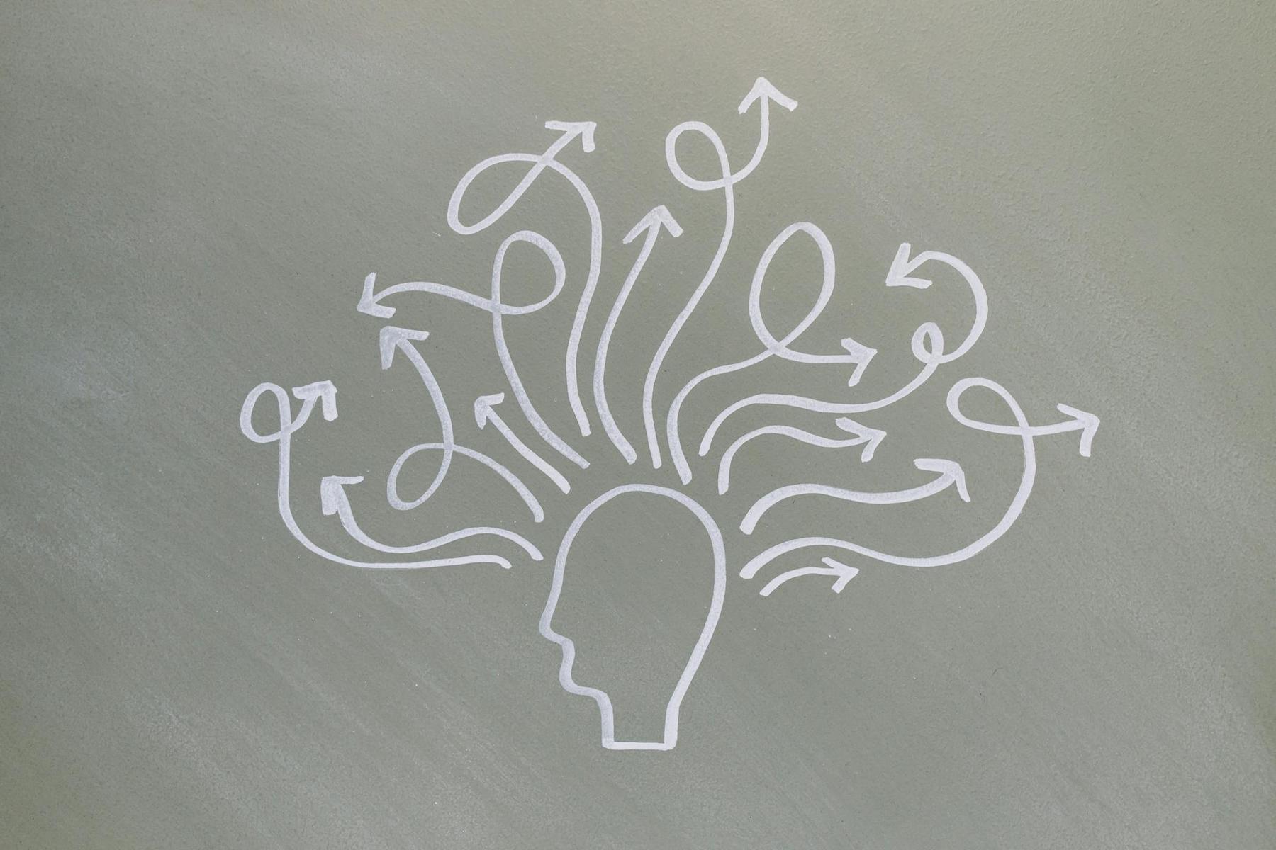
The hypothalamus emerges from this scientific exploration as far more than a simple sleep-wake switch—it represents an exquisitely sensitive integration centre that balances circadian timing, homeostatic need, metabolic status, emotional state, and environmental demands. The suprachiasmatic nucleus serves as master timekeeper, the flip-flop switch ensures stable state transitions, and the orexin system provides stabilising influence that prevents inappropriate sleep intrusions during critical waking activities.
Understanding these mechanisms illuminates why sleep health demands sophisticated, personalised approaches rather than one-size-fits-all solutions. Individual variation in circadian chronotypes, orexin receptor genetics, adenosine sensitivity, and SCN responsiveness to light all contribute to the remarkable diversity in human sleep patterns and requirements.
The Australian context presents unique challenges: extensive seasonal daylight variations across latitudes affect photoperiod and hormonal rhythms differently in tropical Queensland versus temperate Tasmania. Shift work prevalence in mining, healthcare, and emergency services creates widespread circadian misalignment. The 42.9% prevalence of clinically significant sleep disorders in middle-aged Western Australian populations documented in the Raine Study underscores the magnitude of this public health challenge.
Advancing sleep health requires recognition that the hypothalamus operates within broader physiological systems. Light exposure patterns, meal timing, physical activity, stress management, and temperature regulation all influence hypothalamic function. The interaction of multiple signalling systems with the SCN and sleep centres—demonstrating how endogenous regulatory mechanisms show different activation patterns across the sleep-wake cycle—illustrates how multiple physiological systems converge on hypothalamic regulation.
The science of hypothalamic sleep-wake regulation reveals that optimal sleep emerges from alignment—alignment between internal circadian phase and external light cycles, between homeostatic sleep pressure and recovery opportunity, between arousal demands and metabolic capacity. When this alignment breaks down, whether through shift work, sleep disorders, or lifestyle factors, the consequences ripple through every physiological system.
For the 59.4% of Australians regularly experiencing sleep symptoms, understanding the hypothalamic mechanisms underlying these difficulties represents the foundation for addressing them effectively. The remarkable stability of the sleep-wake system—keeping humans in intermediate states less than 2% of each day—demonstrates both the robustness of hypothalamic regulation and the significance of disruptions when they occur.
What makes the hypothalamus so important for sleep regulation?
The hypothalamus contains the suprachiasmatic nucleus (SCN)—the brain’s master circadian clock—along with the ventrolateral preoptic nucleus (VLPO) that promotes sleep and the orexin neurons that stabilise wakefulness. It integrates light information, homeostatic sleep pressure, metabolic status, and hormonal signals to coordinate sleep and wake states. Without proper hypothalamic function, the sleep-wake cycle fragments into unstable episodes rather than consolidated periods of rest and activity.
How does the suprachiasmatic nucleus control the sleep-wake cycle?
The SCN, with approximately 20,000 neurons per side, generates intrinsic 24-hour rhythms via core clock genes like Period, Cryptochrome, Clock, and BMAL1. It receives direct light input through specialised retinal ganglion cells, which reset the clock daily. The SCN then projects to regions such as the subparaventricular zone and dorsomedial nucleus of the hypothalamus, amplifying and distributing circadian signals to sleep-regulatory systems, and also modulates hormonal signalling to help promote sleep.
Can damage to the hypothalamus cause permanent sleep problems?
Yes. Damage to specific hypothalamic nuclei, such as the SCN or VLPO, can severely disrupt circadian rhythms and sleep architecture. Lesions to the SCN fragment the sleep-wake cycle, while damage to the VLPO can reduce both NREM and REM sleep by up to 50-60% with frequent awakenings. Additionally, loss of orexin-producing neurons is linked to narcolepsy type 1, causing severe sleep-wake instability and cataplexy.
Why do shift workers struggle with sleep despite feeling tired?
Shift workers often experience a misalignment between their internal circadian clock, governed by the SCN, and their socially imposed sleep-wake schedules. The SCN continues to send wake-promoting signals at times when shift workers try to sleep, while homeostatic sleep pressure builds during periods of wakefulness. This misalignment prevents the proper consolidation of sleep, leading to reduced total sleep time and poor sleep quality.
What role does the hypothalamus play in sleep disorders affecting Australians?
The hypothalamus is central to the pathophysiology of many sleep disorders. In conditions like insomnia, there is hyperarousal due to overactivity in hypothalamic-pituitary-adrenal pathways and failure of wake-promoting systems to shut down. Circadian rhythm disorders stem from misalignment of the SCN, while narcolepsy arises from the loss of orexin-producing neurons. Even in disorders like obstructive sleep apnoea, disrupted hypothalamic regulation affects metabolic and cardiovascular functions.


